VEGFR-2
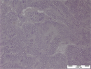
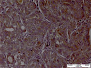
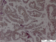
Figure 7(a). Very mild immuno-staining of tumour cells in intratumoral area with VEGFR-2 when treated with citrate AR alone. Original magnification × 1000x.
Figure 7(b). Prominent cytoplasmic and nuclear staining in the intratumoral area with VEGFR-2 when treated with citrate followed by Tris-EDTA AR. Original magnification × 1000x.
Figure 7(c). Marked immuno-reactivity but loss of structural and cellular details of tumour cells with VEGFR-2 when subjected to citrate, followed by Tris-EDTA and pepsin treatment. Original magnification × 1000x.
VEGF-C
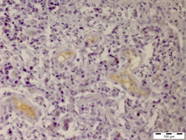
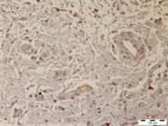
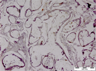
Figure 8(a). Absence of immuno-staining in intratumoral area with VEGF-C when treated with citrate AR alone. Original magnification × 100x.
Figure 8(b). Absence of immuno-staining of in intratumoral area with VEGF-C when treated with citrate followed by Tris-EDTA AR. Original magnification × 100x.
Figure 8(c). Marked loss of cellular details in intratumoral area with VEGF-C when subjected to citrate, followed by Tris-EDTA and pepsin treatment. Original magnification × 400x.